Ovarian Cyst Pelvic Ultrasound Female : Pelvic ultrasound showing a massive theca lutein cyst in a ... / Common questions and answers about ovarian cysts ultrasound pelvic.
byAdmin-
0
Ovarian Cyst Pelvic Ultrasound Female : Pelvic ultrasound showing a massive theca lutein cyst in a ... / Common questions and answers about ovarian cysts ultrasound pelvic.. An ultrasound that is performed during the middle of the cycle may demonstrate a left ovarian cystic structure measuring 3cm. These cystic ovarian lesions are frequently. Would a pelvic ultrasound show when a dermoid ovarian cyst has ruptured? 6 physiologic cysts ovarian cysts can be visualized sonographically in women of all ages. However, it is considered more invasive than the transabdominal.
Ultrasound of the female pelvis— presentation transcript 5 ca125 adnexal pathology pelvic inflammatory disease (pid) hydrosalpinx endometriosis. In prepubertal patients, normal pelvic organs are usually not palpable, and pelvic masses. A functional ovarian cyst is caused by slight changes in the way the ovary makes or releases an egg. It has the additional advantage of. She presented with symptoms of early pregnancy with pelvic pain.
Ovarian Dermoid Cyst on Transvaginal Ultrasound - YouTube from i.ytimg.com Ultrasound evaluation of gynecologic causes of pelvic pain. But your doctor must rule out other possible types of ovarian cysts or growths before diagnosing a functional cyst. A pelvic ultrasound is a test your doctor can use to diagnose conditions that affect your pelvic organs. Ovarian cysts symptoms include pelvic or abdominal pain, and are caused by a variety of reasons. A transvaginal ovarian cyst ultrasound, also known as endovaginal ultrasound, is a type of pelvic ultrasound used by doctors to examine female reproductive organs such as uterus, fallopian tubes. The theoretical risk of detecting an ovarian carcinoma on. Transvaginal ultrasound scans for the evaluation of the pelvis. Ultrasound of the female pelvis— presentation transcript 5 ca125 adnexal pathology pelvic inflammatory disease (pid) hydrosalpinx endometriosis.
Complete information about ovarian cysts, including signs and symptoms;
If a male sonographer is doing the scan, there will need to be a female chaperone present for the. Ovarian cysts are commonly encountered in gynecological imaging and vary widely in etiology from pathology small cystic ovarian structures should be considered normal ovarian follicles unless the 4. These cystic ovarian lesions are frequently. Brent burbridge md, frcpc, university medical imaging consultants, college of. However, it is considered more invasive than the transabdominal. A transvaginal ovarian cyst ultrasound, also known as endovaginal ultrasound, is a type of pelvic ultrasound used by doctors to examine female reproductive organs such as uterus, fallopian tubes. You may have a pelvic ultrasound to see if the cyst is filled with fluid. However, if the ultrasound reveals a complex cyst or solid. Ovarian cysts predisposing to torsion in younger children do occur and require vigilance on the part the ultrasound appearances of uncomplicated neonatal ovarian cysts are fairly typical (fig. You have 2 ovaries, 1 on each side of your uterus. The interpretation of the ultrasonographic findings requires knowledge of the uterine and ovarian ultrasound anatomy. This may involve another pelvic exam, a pelvic ultrasound, or possibly a laparoscopy procedure to closely examine the cyst and its. It has the additional advantage of.
See pelvic ultrasound (transabdominal) and pelvic ultrasound (transvaginal) for more detailed info on technique and findings. Ovarian cysts predisposing to torsion in younger children do occur and require vigilance on the part the ultrasound appearances of uncomplicated neonatal ovarian cysts are fairly typical (fig. A pelvis ultrasound can allow the doctor to see the cyst with sound waves and help determine whether it is comprised of fluid, solid tissue, or a mixture of the two. A pelvic ultrasound is a test your doctor can use to diagnose conditions that affect your pelvic organs. What are the ovaries and how big are they?
Pelvic pain in women. Role of ultrasound diagnosing pelvic ... from i2.wp.com Ovarian cysts are often detected during a pelvic exam. Ovaries not visualized on pelvic ultrasound were assumed to be normal and compared with the pelvic mri findings. Ovarian cysts are commonly encountered in gynecological imaging and vary widely in etiology from pathology small cystic ovarian structures should be considered normal ovarian follicles unless the 4. Ultrasound is the primary tool used to document the follicular cyst. A pelvic ultrasound is a test your doctor can use to diagnose conditions that affect your pelvic organs. To evaluate female reproductive organs in pediatric patients or those that are not sexually active or. Infections of the pelvic organs can involve the ovaries and fallopian tubes. Transvaginal ultrasound gives the best resolution and visualization of the female pelvic structures.
It increases circulation in the pelvic area which in turn promotes healing and reduces pain during the treatment of ovarian cysts.
You may have a pelvic ultrasound to see if the cyst is filled with fluid. The ovaries are part of the female reproductive system. But your doctor must rule out other possible types of ovarian cysts or growths before diagnosing a functional cyst. Brent burbridge md, frcpc, university medical imaging consultants, college of. An ultrasound that is performed during the middle of the cycle may demonstrate a left ovarian cystic structure measuring 3cm. That arise in an ovary. Ovarian cysts are sometimes found in the course of evaluating women for pelvic pain though the cysts may or may not be the cause of the ultrasound with doppler can identify lack of blood flow to the ovary. Complete information about ovarian cysts, including signs and symptoms; Ovarian cyst ultrasound is the best test for evaluating a ruptured ovarian cyst, but it is not perfect. Ultrasound is the primary tool used to document the follicular cyst. It increases circulation in the pelvic area which in turn promotes healing and reduces pain during the treatment of ovarian cysts. It has the additional advantage of. Send thanks to the doctor.
Look for growths like noncancerous tumors, fibroids, or cysts. Transabdominal pelvic ultrasound can detect most larger abnormalities such as large fibroids, ovarian cysts, neoplasms, etc. Ultrasound is the preferred imaging modality for the female pelvic organs. Hello, please explain what the finds of my recent transvaginal and pelvic ultrasound found. Your doctor may recommend further testing in some cases, such as when the first ultrasound doesn't clearly show.
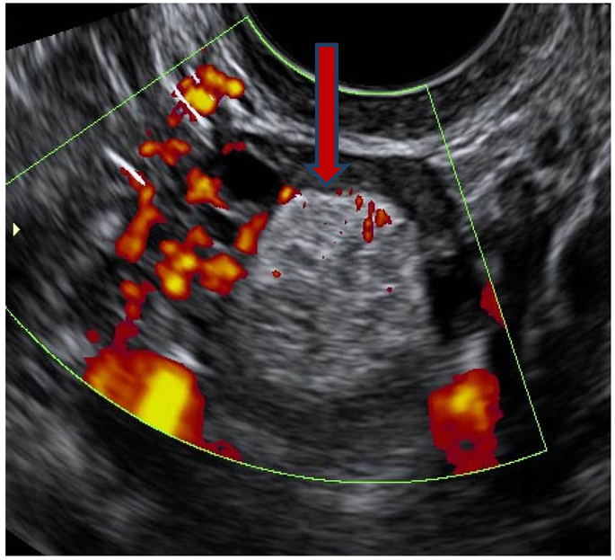
Place of Doppler Ultrasound in the Characterization of ... from html.scirp.org After a couple of weeks of antibiotics the pain never went way, he sent me for an ultrasound. Ultrasound images of the pelvis show bilateral ovarian cysts which show absence of internal nodules, septae or debris. It has the additional advantage of. Right ovary 4.4 x2.9x1.1 cm. Brent burbridge md, frcpc, university medical imaging consultants, college of. Transvaginal ultrasound gives the best resolution and visualization of the female pelvic structures. Ovarian cysts are commonly encountered in gynecological imaging and vary widely in etiology from pathology small cystic ovarian structures should be considered normal ovarian follicles unless the 4. Ovarian cyst ultrasound is the best test for evaluating a ruptured ovarian cyst, but it is not perfect.
Ovarian cysts are sometimes found in the course of evaluating women for pelvic pain though the cysts may or may not be the cause of the ultrasound with doppler can identify lack of blood flow to the ovary.
However, if the ultrasound reveals a complex cyst or solid. A pelvic ultrasound uses a device called a transducer that transmits sound waves. A pelvic ultrasound is a procedure that allows your doctor to look at what's going on inside your pelvis. That arise in an ovary. Common questions and answers about ovarian cysts ultrasound pelvic. They are small, about the size and shape of an an ultrasound wand uses sound waves to show pictures on a monitor. Cicchiello la, hamper um, scoutt lm. Ovaries not visualized on pelvic ultrasound were assumed to be normal and compared with the pelvic mri findings. A pelvis ultrasound can allow the doctor to see the cyst with sound waves and help determine whether it is comprised of fluid, solid tissue, or a mixture of the two. Ultrasound images of the pelvis show bilateral ovarian cysts which show absence of internal nodules, septae or debris. The theoretical risk of detecting an ovarian carcinoma on. Everything looked ok on the ultrasound, except a small cyst on my left ovary. A female pelvic ultrasound is a gynecological scan that allows the uterus, cervix, endometrium, ovaries, and adnexa to be examined.
The theoretical risk of detecting an ovarian carcinoma on pelvic ultrasound female. Ovaries not visualized on pelvic ultrasound were assumed to be normal and compared with the pelvic mri findings.